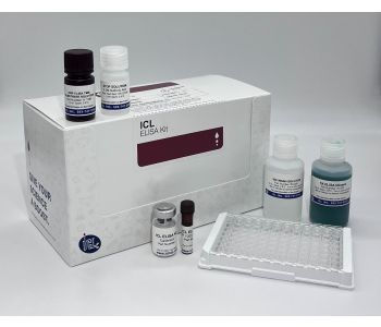
| Antibody Type | ELISA |
|---|---|
| Reactivity | Murine/Mouse |
| Host | Goat |
| Specificity/Target | IgG Fc |
| Size | 1.0 |
| Detection Range | 6.25 ng/ml - 200 ng/ml |
| Sensitivity | 2.374 ng/ml |
| Assay Time | 100 min. |
| Sample Type | Plasma, Serum |
| Storage | 2-8C |
PRINCIPLE OF THE ASSAY
The principle of the double antibody sandwich ELISA is represented in Figure 1. In this assay the IgG present in samples reacts with the anti-IgG antibodies, which have been adsorbed to the surface of polystyrene microtitre wells. After the removal of unbound proteins by washing, anti-IgG antibodies conjugated with horseradish peroxidase (HRP) are added. These enzyme-labeled antibodies form complexes with the previously bound IgG. Following another washing step, the enzyme bound to the immunosorbent is assayed by the addition of a chromogenic substrate, 3,3’,5,5’-tetramethylbenzidine (TMB). The quantity of bound enzyme varies directly with the concentration of IgG in the sample tested; thus, the absorbance, at 450 nm, is a measure of the concentration of IgG in the test sample. The quantity of IgG in the test sample can be interpolated from the standard curve constructed from the standards, and corrected for sample dilution.
Anti-IgG Antibodies Bound To Solid Phase
Standards and Samples Added
IgG * Anti-IgG Complexes Formed
Unbound Sample Proteins Removed
Anti-IgG-HRP Conjugate Added
Anti-IgG-HRP * IgG * Anti-IgG Complexes Formed
Unbound Anti-IgG-HRP Removed
Chromogenic Substrate Added
Determine Bound Enzyme Activity
INTENDED USE
The IgG test kit is a highly sensitive two-site enzyme linked immunoassay (ELISA) for measuring IgG in mouse biological samples. If the ELISA is to be used outside the intended use, the user may need to optimize for said use.
LIMITATION OF THE PROCEDURE
FOR RESEARCH USE ONLY. NOT FOR DIAGNOSTIC PURPOSES. IN VITRO USE ONLY. Reliable and reproducible results will be obtained when the assay procedure is carried out with a complete understanding of the information contained in the package insert instructions and with adherence to good laboratory practice. Factors that might affect the performance of the assay include instrument function, cleanliness of glassware, quality of distilled or deionized water, and accuracy of reagent and sample pipetting, washing technique, incubation time or temperature. Do not mix or substitute reagents with those from other lots or sources.
KIT COMPONENTS
The expiration date for the kit and its components is stated on the box label. All components should be stable up to the expiration date if stored and used per this kit protocol insert.
MATERIALS REQUIRED BUT NOT PROVIDED
- Precision pipettes (2 µL to 100 µL) for making and dispensing dilutions
- Test tubes
- Squirt bottle or Microtitre washer/aspirator
- Distilled or Deionized H2O
- Microtitre Plate reader
- Assorted glassware for the preparation of reagents and buffer solutions
- Centrifuge for sample collection
- Anticoagulant for plasma collection
- Timer
SPECIMEN COLLECTION AND HANDLING
All blood components and biological materials should be handled as potentially hazardous. Follow universal precautions when handling and disposing. If blood samples are clotted, grossly hemolyzed, lipemic, or the integrity of the sample is of concern, make a note and interpret results with caution. The sample collection and storage conditions listed below are intended as general guidelines. Sample stability has not been evaluated.
- Serum samples - Blood should be collected by venipuncture. The serum should be separated from the cells after clot formation by centrifugation. Remove serum and assay immediately or aliquot and store samples at –80°C (preferably) or -20°C. Avoid repeated freeze-thaw cycles.
- Plasma samples - Blood should be collected into a container with an anticoagulant and then centrifuged. Assay immediately or aliquot and store samples at –80C (preferably) or -20°C. Avoid repeated freezethaw cycles.
- Urine samples – Collect mid-stream using sterile or clean urine collector. Centrifuge to remove cell debris. Assay immediately or aliquot and store samples at –80°C (preferably) or -20°C. Avoid repeated freeze-thaw cycles.
- Known interfering substances - Azide and thimerosal at concentrations higher than 0.1% inhibits the enzyme reaction.
DILUTION OF SAMPLES
The assay requires that each test sample be diluted before use. All samples should be assayed in duplicate each time the assay is performed. The recommended dilutions are only suggestions. Dilutions should be based on the expected concentration of the unknown sample such that the diluted sample falls within the dynamic range of the standard curve. If unsure of sample level, a serial dilution with one or two representative samples before running the entire plate is highly recommended.
- Serum samples – Recommended starting dilution is 1/400,000. To prepare a 1/400,000 dilution of a sample, transfer 2 µL of sample to 1,998 µL of 1X diluent. This gives you a 1/1,000 dilution. Next, dilute the 1/1,000 by transferring 2 µL into 798 µL of 1X diluent. This gives you a 1/400,000 dilution. Mix thoroughly each stage.
- Plasma samples – Recommended starting dilution is 1/400,000. To prepare a 1/400,000 dilution of a sample, transfer 2 µL of sample to 1,998 µL of 1X diluent. This gives you a 1/1,000 dilution. Next, dilute E-90GXSPP 4 www.icllab.com
- the 1/1,000 by transferring 2 µL into 798 µL of 1X diluent. This gives you a 1/400,000 dilution. Mix thoroughly each stage.
REAGENT PREPARATION
- Bring all reagents to room temperature (16C to 25C) before use.
- Diluent Concentrate - The Diluent Solution supplied is a 5X Concentrate and must be diluted 1/5 with distilled or deionized water (1 part buffer concentrate, 4 parts dH2O).
- Wash Solution Concentrate - The Wash Solution supplied is a 20X Concentrate and must be diluted 1/20 with distilled or deionized water (1 part buffer concentrate, 19 parts dH2O). Crystal formation in the concentrate may occur when storage temperatures are low. Warming of the concentrate to 30-35C before dilution can dissolve crystals.
- Enzyme-Antibody Conjugate - Calculate the required amount of working conjugate solution for each microtitre plate test strip by adding 10 µL Enzyme-Antibody Conjugate to 990 µL of 1X Diluent for each test strip to be used for testing. Dilute immediately before use and protect from light. Mix uniformly, but gently. Avoid foaming.
- Pre-coated ELISA Micro Plate - Ready to use as supplied. Unseal foil pouch and remove plate from pouch. Remove all strips and wells that will not be used in the assay and place back in pouch and re-seal along with desiccant.
- Mouse IgG Calibrator – Prepare according to the lot specific Certificate of Analysis.
ASSAY PROCEDURE
- All samples and standards should be assayed in duplicates.
- The Standards and the test sample(s) should be loaded into the ELISA wells as quickly as possible to
avoid a shift in OD readings. Using a multichannel pipette would reduce this occurrence.
- Pipette 100 μL of sample (in duplicate) into pre designated wells.
- Incubate the micro titer plate at room temperature for sixty (60 ± 2) minutes. Keep plate covered and level during incubation.
- Following incubation, aspirate the contents of the wells.
- Completely fill each well with appropriately diluted Wash Solution and aspirate. Repeat three times, for a total of four washes. If washing manually: completely fill wells with wash buffer, invert the plate then pour/shake out the contents in a waste container. Follow this by sharply striking the wells on absorbent paper to remove residual buffer. Repeat 3 times for a total of four washes. E-90GXSPP 5 www.icllab.com
- Pipette 100 μL of appropriately diluted Enzyme-Antibody Conjugate to each well. Incubate at room temperature for thirty (30 ± 2) minutes. Keep plate covered in the dark and level during incubation.
- Wash and blot the wells as described in Steps 5/6.
- Pipette 100 μL of TMB Substrate Solution into each well.
- Incubate in the dark at room temperature for precisely ten (10) minutes.
- After ten minutes, add 100 μL of Stop Solution to each well.
- Determine the absorbance (450 nm) of the contents of each well within 30 minutes. Calibrate the plate reader to manufacturer’s specifications.
CALCULATION OF RESULTS
- Subtract the average background value (Average absorbance reading of Standard zero) from the test values for each sample.
- Average the duplicate readings for each standard and use the results to construct a Standard Curve. Construct the standard curve by reducing the data using computer software capable of generating a four parameter logistic curve fit. A second order polynomial (quadratic) or other curve fits may also be used; however, they will be a less precise fit of the data.
- Interpolate test sample values from standard curve. Correct for sera dilution factor to arrive at the IgG concentration in original samples.
Manufactured by: Immunology Consultants Laboratory, Inc. (ICL)
This document contains information that is proprietary to Immunology Consultants Laboratory. The original recipient of this document may duplicate this document in whole or in part for internal business purposes only, provided that this entire notice appears in all copies. In duplicating any part of this document, the recipient agrees to make every reasonable effort to prevent the unauthorized use and distribution of the proprietary information.
Immunology Consultants Laboratory, Inc. (ICL) does not provide any type of warranty relating to either product
fitness for application or level of standard, implied or expressed beyond the option of replacing material proven to
be defective. ICL will not be held accountable for expenses or loses incurred from the use of this product. The
buyer bears full responsibility for the use of the product to ensure that the product is suitable for use and to plan
for sufficient quantity
