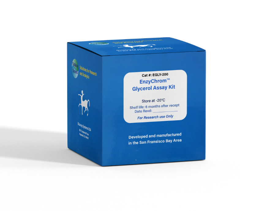
DESCRIPTION
GLYCEROL [GLYCERIN or GLYCERINE, C3H5(OH)3] is widely used in foods, beverages and pharmaceutical formulations. It is also a main byproduct of biodiesel production. Simple, direct and automation-ready procedures for measuring glycerol concentrations find wide applications. BioAssay Systems' glycerol assay uses a single Working Reagent that combines glycerol kinase, glycerol phosphate oxidase and color reactions in one step. The color intensity of the reaction product at 570nm or fluorescence intensity at λem/ex = 585/530nm is directly proportional to glycerol concentration in the sample.
KEY FEATURES
Sensitive and accurate. Use as little as 10 µL samples. Linear detection range in 96-well plate: 10 to 1000 µM (92 µg/dL to 9.2 mg/dL) glycerol for colorimetric assays and 2 to 50 µM for fluorimetric assays.
Simple and convenient. The procedure involves addition of a single working reagent and incubation for 20 min at room temperature, compatible for HTS assays.
Improved reagent stability. The optimized formulation has greatly enhanced the reagent and signal stability.
APPLICATIONS:
Direct Assays: glycerol in biological samples (e.g. serum and plasma).
Drug Discovery/Pharmacology: effects of drugs on glycerol metabolism.
Food and Beverages: glycerol in food, beverages, pharmaceutical formulations etc.
KIT CONTENTS
Assay Buffer: 24 mL Enzyme Mix: 500 µL
ATP: 250 µL Dye Reagent: 220 µL
Standard: 100 µL 100 mM Glycerol
Storage conditions. The kit is shipped on ice. Store all components at -20°C. Shelf life of 12 months after receipt.
Precautions: reagents are for research use only. Normal precautions for laboratory reagents should be exercised while using the reagents. Please refer to Material Safety Data Sheet for detailed information.
COLORIMETRIC 96-WELL PROCEDURE
Note: SH-group containing reagents (e.g. mercaptoethanol, DTT) may interfere with this assay and should be avoided in sample preparation.
1. Equilibrate all components to room temperature. Keep thawed Enzyme Mix in a refrigerator or on ice. Dilute standard in distilled water as follows (diluted standards can be used for future assays when stored refrigerated).
Transfer 10 µL standards and 10 µL samples into separate wells of a clear 96-well plate.
2. For each reaction well, mix 100 µL Assay Buffer, 2 µL Enzyme Mix, 1 µL ATP and 1 µL Dye Reagent in a clean tube. This Working Reagent should be used on the same day of preparation. Transfer 100 µL Working Reagent into each reaction well. Tap plate to mix.
3. Incubate 20 min at room temperature. Read optical density at 570nm (550-585nm).
Note: if the Sample OD is higher than the Standard OD at 1.0 mM, dilute sample in water and repeat the assay. Multiply result by the dilution factor.
CALCULATION
Subtract blank OD (water, #4) from the standard OD values and plot the OD against standard concentrations. Determine the slope using linear regression fitting. The glycerol concentration of Sample is calculated as
FLUORIMETRIC 96-WELL PROCEDURE
For fluorimetric assays, the linear detection range is 2 to 50 µM glycerol. Mix 10 µL 100 mM Standard with 990 µL H2O (final 1 mM).
Dilute standards as above. Transfer 10 µL standards and 10 µL samples into separate wells of a black 96-well plate.
Add 100 µL Working Reagent (see Colorimetric Procedure). Tap plate to mix.
Incubate 20 min at room temperature and read fluorescence at λex = 530nm and λem = 585nm.
The glycerol concentration of Sample is calculated as
MATERIALS REQUIRED, BUT NOT PROVIDED
Pipeting devices, centrifuge tubes, Clear flat-bottom 96-well plates, black 96-well plates (e.g. Corning Costar) and plate reader.
PUBLICATIONS
1. Li, C et al (2020). Matrix Gla protein regulates adipogenesis and is serum marker of visceral adiposity. Adipocyte, 9(1), 68-76.
2. Fontvieille, A et al (2019). Acute effect of post-resistance exercise milk-based supplement on substrate oxidation and fat mobilization in older men: A pilot study. Advances in Geriatric Medicine and Research, 1(2).
3. Pan, Y., et al. (2018). Salvianolic acid B improves mitochondrial function in 3T3-L1 adipocytes through a pathway involving PPARgamma coactivator-1alpha (PGC-1alpha). Frontiers in pharmacology, 9:671.
